| 商品編號: | NEPA21 |
| 商品名稱: | 4-step Super Electroporator 四階超級電穿孔儀 |
| 品牌廠牌: | NepaGene |
| 商品特點: | 高轉殖效率, 高存活率, 應用範圍廣, 無鎖定耗材緩衝液 in vitro, in vivo, ex vivo, in utero and in ovo |
| 檔案下載: | NEPA21 brochure |
NEPA21
4-step Super Electroporator 四階超級電穿孔儀
Ø High transfection efficiency
Ø High viability
Ø No special buffer required
專治 Primary cell, Neuron, stem cell, immune cell 等難搞的細胞
可在懸浮狀態 (cuvette) 或者貼附狀態 (culture dish)進行轉染.
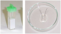
NEPA21 cuvette electroporation
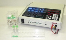
不需要綁定耗材及特定buffer套組, 針對難搞的細胞 ex. primary cells,
stem cells, immune cells, blood cells等, 都可達到高轉殖效率與高存
活率.
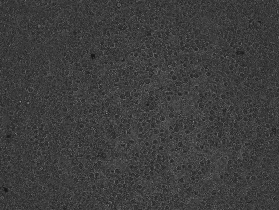
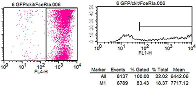
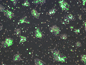
初代細胞轉染
 |
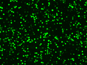 |
| 存活率: 80% | 轉染效率: 83% |
 |
Stem cells, ES cells 及 iPS cells的轉染
 |
| 電穿孔後3天 |
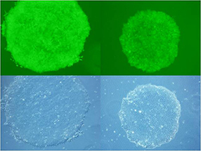 |
| 穩定表現 |
 |
 |
| 存活率: 90% | 轉染效率: 75% |
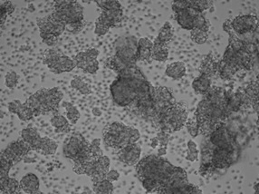
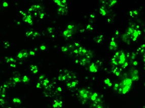
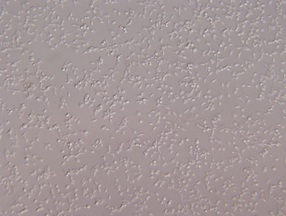
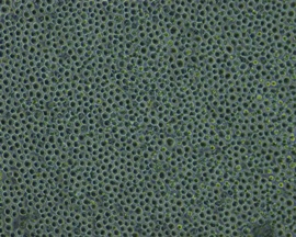
Cell lines的轉染
 |
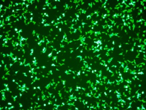 |
| 存活率: 83% | 轉染效率: 87% |
 |
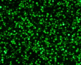 |
| 存活率: 90% | 轉染效率: 85% |
耗材試劑的比較
| 電穿孔設備 | NEPA21 (Nepa Gene) | N (Company L) | N (Company I) |
| 耗材kit |
Cuvette ONLY No Special Buffers |
Transfection Kits with Special Buffers |
Transfection Kits with Special Buffers |
|
處理每個sample 的費用 |
NT$80 | NT$720 ~ 1000 | NT$640 ~ 820 |
您還在忍受昂貴耗材的困擾嗎?
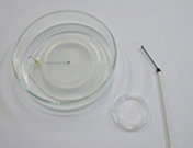
針對貼附型細胞, NEPA21可以直接在24-well, 12-well 或 6-well培養盤進行DNA/RNA電穿孔傳送.
|
|
|
|
|
|
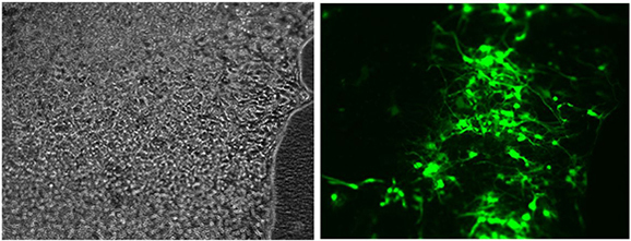
pCAGGS-EGFP plasmid was transferred into primary neurons cultured for 6 days in adherent state.
| A. | 使用貼附細胞用電極 CUY900-13-3-5, 藉由四階超級電穿孔儀來進行轉染 |
| B. | Neuron在電傳送2天後的 EGFP 螢光影像 |
| C. | 圖B的放大圖, 高轉染效率, 可觀察到很多強的ECFG訊號 |
| D. | 圖C的40倍放大圖, 可清楚看到神經軸突(Neurites) |
Data provided courtesy of Department of Neurochemistry, National Institute of Neuroscience, Japan
NEPA21活體電穿孔應用 In Vivo Electroporation
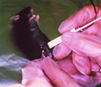 |
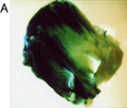
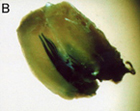
將 pCAGGS-lacZ報導基因以電穿孔方式導入小鼠肌肉. 在基因電傳送5天後以X-gal染色方式觀察LacZ的表現. 可看到在 muscle fibers有大量表現, 同時在control組 (無電穿孔)並沒有表現. |
 |
 |
 |
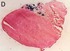 |
| Whole muscle(有電) | Whole muscle(沒電) | 橫切面 (有電) | 橫切面 (沒電) |
 |
 |
 |
 |
 |
 |
| Mouse Brain | Mouse Skin | Mouse Testes | Rat Retina | Honeybee Brain | Silkworm Eggs |
NEPA21電穿孔儀應用 In Utero Electroporation
可以將 DNA/RNA 電傳送到老鼠胚胎 (cerebral cortex, hippocampus, spinal cord, and more.)
 |
| pCAG-EGFP was injected into the both lateral ventricles of E14.5 mouse embryos and electronic pulses (33V, 50msec) were charged four times. 3 days later, the embryos (E17.5) were fixed and the brains were removed and examined under a fluorescence stereomicroscope (Fig. A). Fluorescence was observed in the lateral region of the hemisphere onto which the anode had been placed and in the medial region of the opposite hemisphere. And brains were frozen and sliced and the fluorescent image was obtained with a confocal laser microscope (Fig. B). |
NEPA21電穿孔儀應用 In Ovo Electroporation
 |
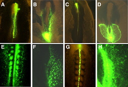 |
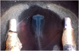
| ransfection into the central canal | Transfection into the somites (A, E), hematopoietic system (B, F), notochord (C, G), and lateral plate mesoderm (D, H) |
NEPA21電穿孔儀應用 Ex Vivo Electroporation
 |
 |
 |
 |
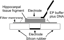
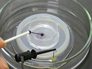
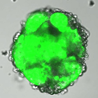
| The neurons were prepared from P7 rat hippocampus. Electroporation was performed after 11 DIV. |
 |
 |
訊詢單內容